Within the field of microscopy, there are several laboratories linked to the Faculty of Science and Technology at UiS. An overview of equipment and facilities follows.
These study programmes use the laboratories in teaching:

Scanning electron microscope (SEM)
The scanning electron microscope is part of the material characterisation activities at the faculty. Samples can be imaged down to micro and nanometre levels. This is possible because electron microscopes use electrons to illuminate the sample/create a magnified image of what is being analysed, unlike ordinary optical microscopes that use light.

Bombarding a sample with electrons produces various signals that provide information about the surface, microstructure, and chemical composition. Users can take images using secondary electrons (SE), backscattered electrons (BSE) and cathodoluminescence (CL), as well chemical analysis using energy dispersive X-ray spectroscopy (EDS) and crystal orientation mapping by electron backscattered diffraction (EBSD). Numerous disciplines use SEM and the equipment is available for use by students, staff, and external stakeholders by agreement.
Contact person: Espen Undheim
Transmission electron microscopy (TEM)
Transmission electron microscopy (TEM) is a microscopy technique in which a beam of electrons is transmitted through a sample to form an image or diffraction pattern to characterize various microstructural and crystallographic properties of materials.
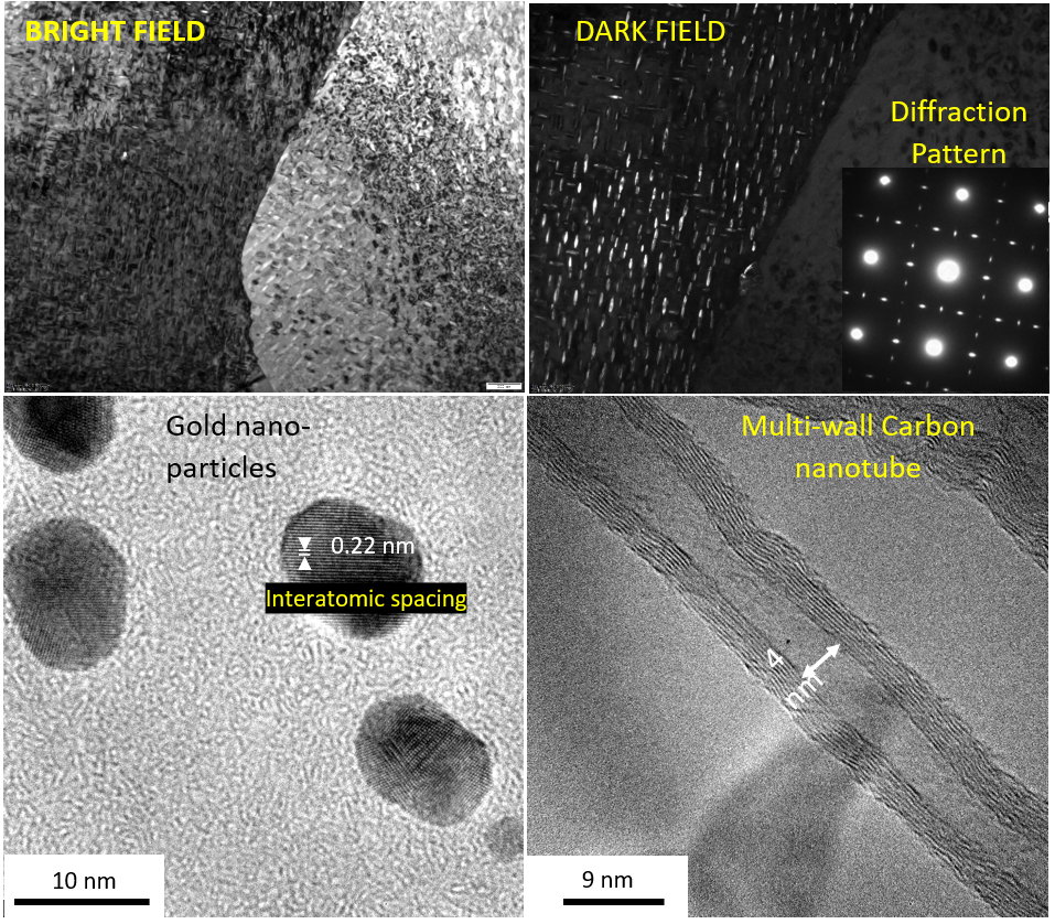
Primarily, TEM is used to analyse morphology, composition, crystal defects and phases of materials. Because of the small wavelength effect of the electrons, TEM provides a resolution down to an atomic level. In principle, all kinds of substances can be studied by TEM. For analysis with conventional TEM, the samples must be electron transparent and conductive. The TEM is used by students and staff as well as external research institutions and industry.
Contact person: Wakshum Mekonnen
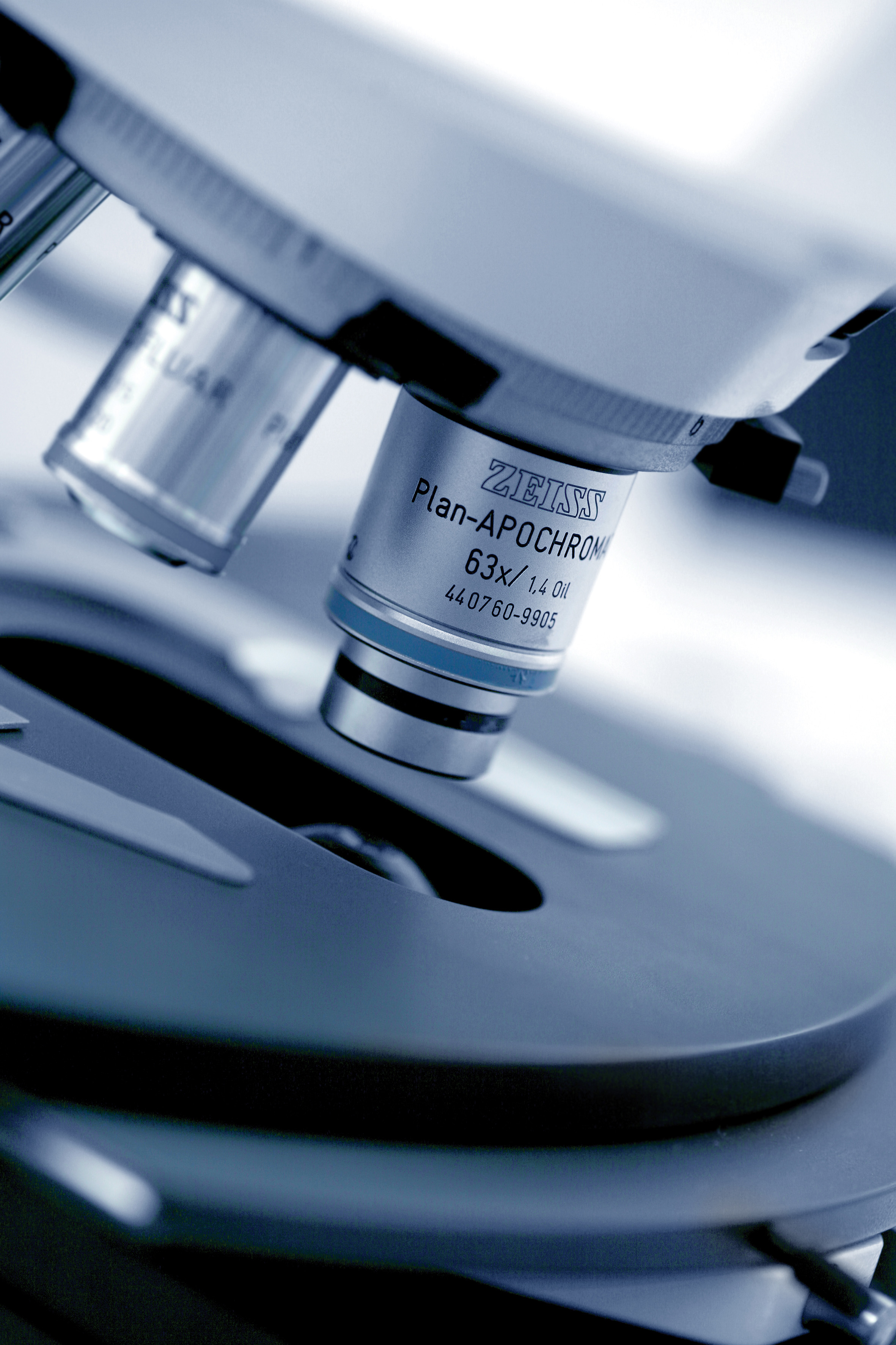
Light microscopy
The Faculty of Science and Technology has many different types of microscopes, which are used in laboratories across multiple disciplines. These microscopes include stereo microscopes, inverted microscopes and polarisation microscopes with different magnification options. Several of the microscopes have associated cameras and software for image processing.
Contact persons: Caroline Ruud (geology), Julie Nikolaisen (Chemistry, environmental engineering, biology), Johan Andreas Håland Thorkaas (metallurgy,inverted light microscopes).
Raman spectroscopy
With the help of Raman spectroscopy, the structure of materials can be examined. The technique is based on the Raman effect and is based on molecular oscillations or lattice oscillations that are generated when a substance is exposed to electromagnetic radiation
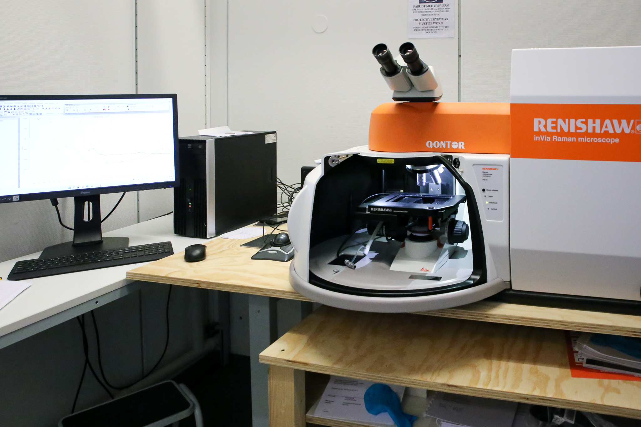
In Raman spectroscopy a laser is applied to irradiate samples. This will polarise and excite the molecules, leading to frequency-shifted scattering, which carries information about the vibration frequency of the molecule in question. From the measured vibration spectra, one may learn about the binding, symmetry, and structure of molecules.
Contact person: Olena Zavorotynska
Cathodoluminescence microscopy
A cathodoluminescence microscope combines the methods of an electron microscope with the methods of a regular light-optical microscope. This makes it possible to study structures in crystals or substances that cannot be seen under normal lighting conditions.
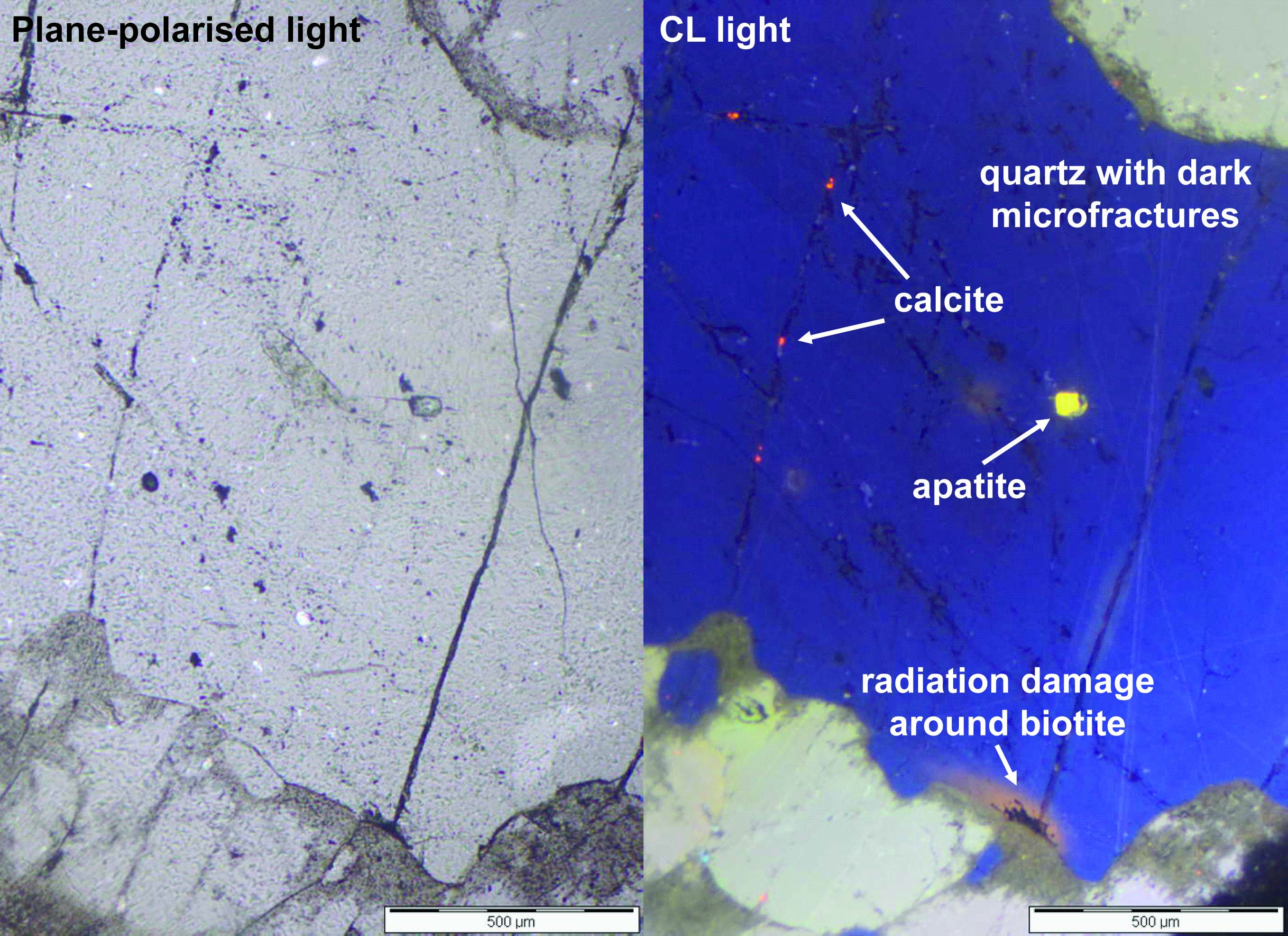
Cathodoluminescence is a light effect on many minerals that occurs when the mineral is bombarded with electrons. The more impurities and crystal-lattice defects the material has, the more likely it is to give a cathodoluminescence signal. In the cathodoluminescence microscope, the effect can be seen as light in different colours, and the light can be measured for its wavelength spectrum.
Contact persons: Carita Augustsson and Caroline Ruud
Confocal microscopy
Confocal microscopy is used to visualize subcellular structures, as you are able to focus a small beam of light at a narrow depth level at a time. Confocal microscopy is an optical imaging technique for increasing optical resolution by means of using a spatial pinhole to block out-of-focus light in image formation.
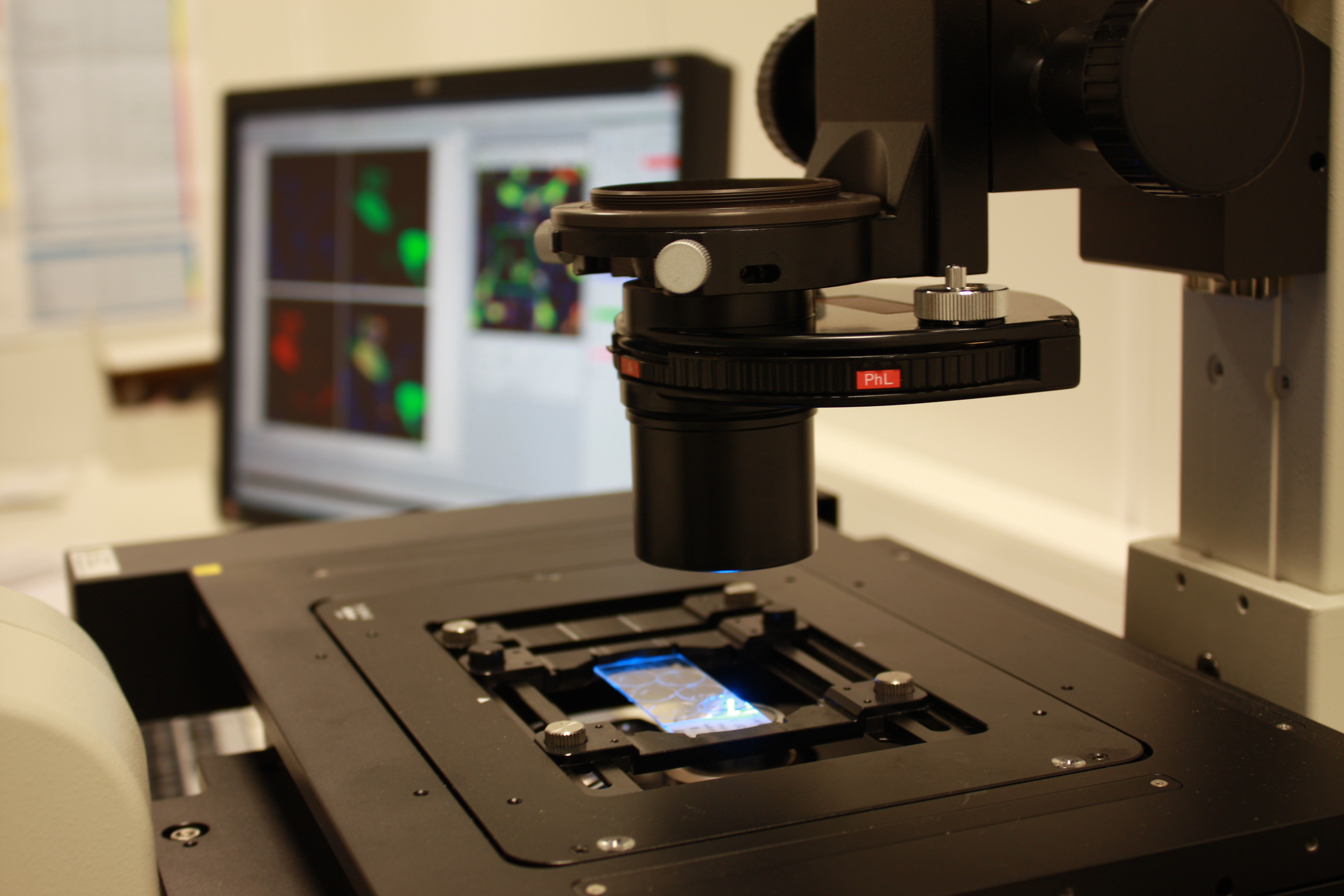
Capturing multiple two-dimensional images (x,y plane) at different depths (z plane) in a sample enables the reconstruction of three-dimensional structures (a process known as optical sectioning) within an object. This is commonly used in visualizing structures within life sciences but may also be used for materials sciences.
Contact person: Hong Lin
You might also be interested in:

Environmental Neuroscience
Environmental neuroscience integrates environmental, social, and cognitive psychology with social, cognitive, and behavi...

12 million for research in algebraic geometry
Professor Helge Ruddat has received NOK 12 million to investigate and classify the mathematical concept of Fano manifold...

HyTack: Tackling the Challenges in Hydrogen Economy through Education and Research
The HyTack project aims at developing and providing an educational base in the field of hydrogen technology to the stude...

New evidence for quark matter cores in massive neutron stars
Researchers at the University of Stavanger are now one step closer to finding out what is in the core of neutron stars. ...

Data-driven mathematical modelling
Mathematical models enable a scientific understanding of natural phenomena around us, and the study and optimization of ...

Whole Slide Imaging, Digitalization and Automation of Histopathological Lesion Evaluation in Marine Organisms
Use of histopathology to identify changes in marine species in response to contaminants is a long-known practice.
Geometry and analysis
The group conducts research in algebraic geometry, complex analysis and geometry, mathematical models and differential e...

Laboratories for chemistry and environmental technology
The Faculty of Science and Technology at UiS has several laboratories within the field of chemistry and environmental te...

Can blueberries prevent dementia?
Drinking juice with a lot of antioxidants, for example from blueberries, can be beneficial in preventing dementia. New r...

Laboratories for materials science
The Faculty of Science and Technology at UiS has several laboratories within the field of materials science. Here is an ...

Laboratories for biology and molecular biology
The Faculty of Science and Technology at UiS has several laboratories within the field of biology and molecular biology....

Functional materials and process chemistry group
We are developing functional materials and process chemistry for energy and environmental applications.

Library resources: Science and Technology
Do you need help finding literature for your assignment or thesis? Or help getting started with referencing? Check out o...

UiS partner in new One Health project
The purpose of the new research project is to support advancement of Blastocystis research by bringing together professi...

In the core of a neutron star
How can the established theory of particle physics, the Standard Model, be used to predict the material properties of th...

SØLSTAIN: Red seaweed reproduction, growth and nutritional quality
Our research aims to contribute to a sustainable use of the red seaweed Palmaria palmata in aquaculture. The work focuse...

Sanocean
Marine Sewage Outfalls – Environmental Impact Evaluation (SANOCEAN) focusing on ocean research including blue economy, c...

Porous Liquids: New porous liquids for gas separation and carbon capture
The Porous Liquids project aims at developing a new technology for environmentally friendly and efficient separation of ...

Funding for new research project on intestinal diseases
University of Stavanger (UiS) and Stavanger University Hospital (SUS) will work together to find solutions for patients ...

MOFsorbMET: Metal-organic frameworks for recovery and separation of critical metals
The MOFSORBMET project is developing MOFs for the adsorption-enhanced recovery of critical metals from the primary (mini...